Diagnosis is necessary to draw up a high-quality treatment plan that will maximise results. Only a professionally organized set of measures can help a competent specialist to make the correct diagnosis and provide adequate treatment.


“Dental Club” performs 3D diagnostics using the latest generation equipment.
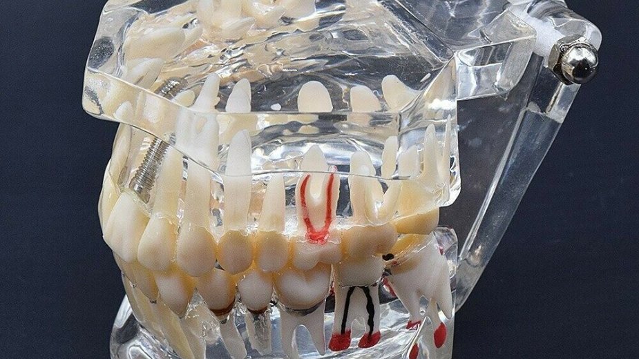
The Planmeca ProMax 3DMid CT scanner is one of the world's best diagnostic devices. ProMax 3DMid is a guarantee of safety. The level of radiation during the examination does not exceed the level of radiation acquired after a short air flight. Thanks to the capabilities of this device, it is possible to create a virtual model of a patient’s teeth and bone tissue.
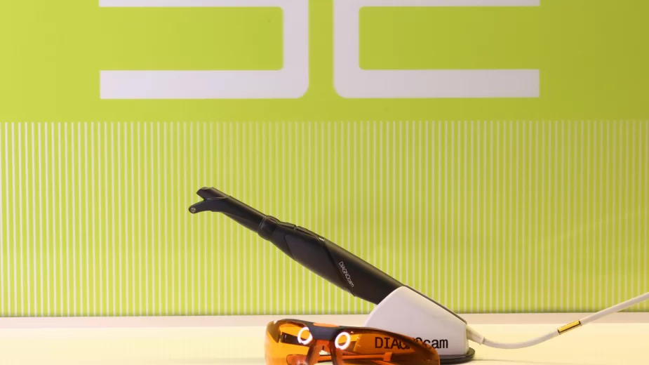
DiagnoCam (KaVo, Germany) is the second tool in the “Dental Club”, used to detect the smallest defects and hidden decay cavities. DiagnoCam uses a laser diagnostic system, but without radiation. The patient can watch the entire diagnostic process on a monitor screen.
“Dental Club” specialists always perform examinations using special optical magnifiers: Leica dental microscope and Carl Zeiss binoculars. The accuracy of the diagnosis is proportional to the magnification coefficient of these devices - ten times more accurate than with a simple examination.
Our team consists of professionals who annually improve their knowledge in leading medical institutions in Europe.


Each patient will have their own electronic file that allows us to track all stages of their treatment.
Stages
- Questionnaire and survey.
- Examination of the patient in a comfortable environment, using Carl Zeiss binoculars or a Leica microscope.
- If necessary, visualization of hidden cavities and microdefects of teeth with DiagnoCam.
- 3D digital diagnostics, with a huge range of functionality: from local areas of teeth, joints and jaws, to all bone tissues of the head.
- Discussion of the results with the patient and joint treatment planning.

Check the actual cost by this number: 87073138812
| CBCT (3D image) | 30 000 KZT |
| OPG (2D image) | 8 500 KZT |
| Diagnocam (laser diagnostics) | 10 000 KZT |
| Leica microscope | up to 30,000 KZT |
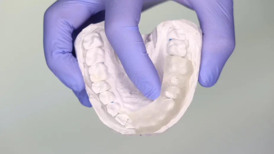
| Scanning with Planmeca intra-oral scanner (1 jaw) | 20 000 KZT |
For a day of life with a visit to the beach, we get 22 μSv; 40 μSv is a few hours of flight by plane, and in a year we "eat" about 400 μSv with food. But it is not recommended to abuse X-rays, more than 3 times a month.
It is not recommended to take process an X-ray during the first 18 weeks. After 18 weeks, it will be more correct to take a two-dimensional or targeted x-ray image. In the event of a situation that requires an X-ray during pregnancy, it is important to remember that the radiation dose of the child is 1/50000 of the mother's dose for an X-ray of the head, this amount is considered safe.
Detection of cavities in their entirety: from initial forms to extensive lesions.
Real-time images (visualization of the diagnostic process on the screen).
The absence of X-ray radiation makes it possible to detect changes in patients with radiophobia, cancer patients, children, pregnant women and sedentary patients.
A microscope can magnify the object by 40 times, correspondingly, the quality of diagnosis and treatment increases significantly.
Binoculars magnify the image by 5 times and are a more compact device used in everyday work.